Resolving the causes of pain. What is the idea behind it?
"Pain is the tissue's cry for flowing energy." This insight is attributed to Huángdi Néijing, who set it down as early as approximately 2500 BC in *The Yellow Emperor's Medicine*. And this experience is likewise expressed in the saying regarding the ailments of the elderly: "If you wake up in the morning without pain, you died during the night."
Why do these insights arise in the context of fascia therapy? The focus of this chapter lies on the body's electrical nature and therapy utilizing bio-analogous microcurrents. The body's natural electrical energy is crucial to both life and death; after all, we are not considered clinically dead until our brainwaves cease. Yet, outside the fields of neurology and cardiology, this electrical vitality receives scant attention. This neglect is unjustified, for every electrical signal—originating from nociceptors (pain receptors) and traveling to the brain—is interpreted there as pain. These receptors respond to the pressure of a crushing injury, the heat of a burn, or the corrosive effect of an acid. The objective behind this sensation of discomfort is clear: we seek to identify and resolve the underlying causes as quickly as possible. We instinctively pull our fingers out of a door, snatch our hand away from a hot stove, or wash off corrosive acid. But what are we to do when we carry that "acid" deep within our own tissues? This localized acidification is a widespread phenomenon and is considered the root cause of approximately 90% of all chronic pain. It is the source of those complaints—often classified as "nonspecific"—for which no underlying cause can be identified despite extensive diagnostic testing. It manifests as rock-hard muscles, chronically adhered fascia, and restricted mobility! Almost invariably, alongside these acid accumulations, there is yet another factor contributing to persistent, agonizing pain. This factor explains why the intense pain of a crushing injury does not immediately subside completely the moment we pull our finger out of the door.
The initially sharp, sudden impulse does not vanish; it merely changes its quality, transforming into a dull, throbbing sensation. First, it serves as a warning signal, prompting us to remove ourselves from danger. Then, it becomes a discomfort—one we could really do without. Theory suggests that the nociceptors (pain receptors) have incurred an "oxygen debt" during the transmission of electrical signals. This dull pain signals this oxygen deficit until the debt has been repaid. In this scenario, the tissue is crying out for oxygen—oxygen that must diffuse from the microcirculation, through the extracellular fluid, to the cells.
Tissue damage resulting from such an injury inevitably leads to swelling. Just consider a sprain (distortion) of the ankle: a distinct lump quickly forms over the overstretched ligament. This local fluid accumulation creates a longer diffusion distance between the capillaries and the tissue cells. Consequently, oxygen reaches the free nerve endings more sparsely, and we are forced to endure the dull, aching pain for a significantly longer period. Furthermore, repair processes are delayed because fewer building blocks are delivered to the site.
Nach Darwin setzt sich in der Evolution nur das Positive durch. Wieso According to Darwin, only the positive prevails in evolution. Why did this swelling—which delays healing—not end in a dead end, but instead persist in both humans and animals?
It is likely indispensable in initiating the body's self-healing mechanisms. How so? Our bodily fluids transport electrolytes and ions into the swollen tissue. As a result, the normal electrical resistance—measured in ohms—is locally reduced. Measurements taken on an inflamed kidney revealed a significantly stronger electric field than that found in a healthy kidney. From the perspective of microcurrent therapists, this process marks the onset of self-healing. What purpose, then, does this process serve?
Let us return to the example of a sprain. Healthy cellular tissues exhibit a uniform polarity within their cell membranes. Injured structures, conversely, display a chaotic distribution of positively and negatively polarized fields. As long as this state persists, organized wound healing cannot commence. By lowering electrical resistance, a significantly greater amount of the body's own energy can exert a regulatory influence. Only when the primary task—restoring the normal potential across the cell membranes—has been completed does the swelling subside and structural repair begin. One key factor contributing to the immense benefit of stimulating healing with microcurrent devices is this: the externally applied, bio-analogous electricity rapidly restores this uniform potential. Tissue healing therefore begins much sooner. Pain levels are significantly reduced as early as the very first session. A study published in February 1990 by H. Lathrop, PhD, in the *Worker's Comp Advisor*, confirms that microcurrent therapy reduces the healing time for acute injuries—sustained by patients in mining-related workplace accidents—by half.
What is impressive about this study is that three large groups—each consisting of 250 patients—were compared. Group I received only microcurrent therapy in the microampere range (millionths of an ampere); Group II received a pharmaceutical agent in addition to microcurrent therapy; and Group III received the same medication combined with a much stronger form of electrical stimulation therapy in the milliampere range (thousandths of an ampere). The result: On average, participants in Groups I and II were fit to return to work after fewer than nine days of therapy. As a user of microcurrent therapy, you can therefore confidently forgo the costs and side effects associated with pharmaceutical medications. On average, participants in Group III required more than twice as many days of therapy. These studies clearly demonstrate the healing effects of microcurrent therapy. Have you previously been skeptical of electrotherapy? You were likely only familiar with stimulation current or TENS methods operating in the milliampere range.
IMPORTANT: For a microcurrent treatment, the practitioner calculates the intensity based on the body's own electrical properties: five millionths of an ampere are required per square centimeter of electrode surface area. The tissues respond immediately, as they are stimulated not only by energies analogous to those of the body but also by proven frequencies.
Frequency therapy is defined by the fact that energy is not conducted through the tissue between the electrodes as a continuous galvanic direct current—as it would be from a battery. Instead, the gentle flow of energy pulses on and off rhythmically. The unit of measurement for this is "Hertz." When set to one Hertz, current flows once every second for a duration of half a second, followed by a half-second interruption. When regulating cells using high-quality microcurrent devices, two galvanically isolated channels simultaneously deliver different frequencies into the pain zone. Although both channels are housed within a single unit, you should visualize it as if two separate devices were simultaneously directing their therapeutic currents into the body. This approach—known as frequency-specific microcurrent therapy—is based on the understanding that specific frequencies (or rhythms) exist that resonate with only one particular biological structure. In this context, "resonance" means that the specific tissue begins to vibrate in sync with the frequency. The benefit is that while all tissue types receive energy, only that one specialized structure derives the maximum therapeutic benefit. You might visualize this much like a soccer match: all the players are in motion, yet only one is moving the most—the player currently in possession of the ball! In the context of established microcurrent therapy, this corresponds to the specific tissue that enters into resonance. For instance: if one channel delivers stimulation at 10 Hertz, the spinal cord will resonate with it. Simultaneously, the second channel will deliver a sequence of distinct frequencies to this now-resonant structure, thereby assigning it specific therapeutic tasks. Continuing our example: if the second channel stimulates at 49 Hertz, the vitality of the spinal cord is enhanced. It is akin to a language, or like the use of "bush drums" in Africa: anyone capable of interpreting the rhythmic drumbeats immediately understands the message—for instance, "Our gathering is about to begin!" Additionally, careful consideration must be given to the proper placement of the adhesive electrodes, as this determines and directs the specific pathway of the energy flow through the body. Frequency-specific microcurrent therapy recommends positioning the electrodes such that the imaginary line connecting the cathode and anode passes directly through the pain zone. In our example of a sprained ankle, the electrode pair corresponding to the tissue frequency could be placed on the inner and outer sides of the affected joint. This placement interacts with the influence of the other electrode pair, which is positioned either front and back or top and bottom. Both frequencies intersect within the painful area, establishing what is known as an interference field. In this process, the applied rhythms undergo both addition and subtraction. It is akin to listening to two tribal drums of identical timbre, sounding simultaneously—each following its own distinct, fixed rhythm. You no longer perceive 49 and 10 Hertz as separate entities, but rather a unique, blended composition.
**The Unique Feature:** Presumably, the cellular structure does not recognize that this therapeutic vibration is acting upon it from the outside. It presents itself as if it were endogenous information—information originating from the body itself. We should move away from the maxim that "more is better." Figuratively speaking, anyone who "screams" at diseased cells using high-intensity stimulation current actually drives the transport activity of the cell membranes down to 60%. Instead, we should "speak softly and kindly," which causes membrane transport to rise to 140%. The result of this improved supply is a 170% increase in protein synthesis—a process essential for any form of cellular repair!
These impressive improvements were published by the team of Cheng et al. in 1989 in *Clinical Orthopaedics* (USA). Also noteworthy is the simultaneously measured increase in cellular energy—specifically Adenosine Triphosphate (ATP)—which rose to five times its baseline level. The fact that conventional stimulation current therapy was measured to cause a decline in membrane transport, protein synthesis, and ATP levels—dropping them to roughly half their normal values—explains why acute injuries heal in half the usual time when treated with microcurrent therapy.
** Oxygen Deficiency as a Cause of Pain **
As previously mentioned, oxygen deficiency is a significant cause of pain. When extremely severe—as is the case during a heart attack—it can even lead to what is known as "crushing pain." Let us recall the wisdom of early Chinese medicine cited at the beginning of this text. During a heart attack, blood flow through the coronary arteries is obstructed. The pain is, in essence, a desperate cry for increased blood flow—and, consequently, for oxygen.
In addition to arterial circulatory disorders, chronic pain can also be triggered by an oxygen deficiency resulting from prolonged muscular tension or rigidity. Thickened myofibrils (muscle fibers) constrict the lumen (cross-section) of the vessels supplying them. A well-known consequence of an imbalance between oxygen demand and supply is a metabolic shift toward energy production via glucose utilization. The resulting lactic acid accumulates locally within the soft connective and supporting tissues. This pH shift has been impressively confirmed through comparative measurements taken between trigger points (registering only pH 4.5) and normal muscle tissue (pH 6.5). Every massage therapist recognizes these localized "acid pools"—palpable around trigger points—as myogeloses.
Fasciae Are Fascinating
We owe the realization that the elastic connective tissue linings of the bile ducts are not—as previously assumed—rigid plates to studies utilizing laser light imaging. On March 28, 2018, a research team led by Dr. Neil Theise at the New York University School of Medicine published findings in *Scientific Reports* demonstrating that all mobile structures are enveloped by a familiar network composed of sturdy collagen and flexible elastin proteins. The key insight: the extracellular matrix stores its fluid within sponge-like compartments formed by this very network. This explains the protective—and, above all, the shock-absorbing—properties of these tissues. Furthermore, this structure ensures that fasciae—thanks to the embedded ion-rich fluid—respond effectively even to gentle microcurrent therapies.
A fascinating aspect of elastin is the fact that it is synthesized only during the prenatal period and the first few years following birth; thereafter, its production virtually ceases. With a half-life of 74 years, it is an exceptionally durable substance—a longevity explained by its extensive resistance to proteolytic (protein-degrading) enzymes. One might assume, then, that everything is in order.
Yet, Fasciae Remain Vulnerable
And yet, adhesions and damage to these remarkably durable structures occur time and again. They lose their elasticity—for instance, the plantar fasciae in the soles of the feet can become extremely sensitive to pressure and as hard as a board. Granted, few people enjoy walking barefoot over pebbles, but that might not seem like a truly serious problem just yet.
Far more significant, however, is the loss of elasticity in the arterial walls. Consequently, the pulse wave generated by each heartbeat is no longer propelled forward with the same vigor as it was in one's youth. Cardiovascular diseases rank among the leading causes of death in the industrialized world.
A realization that has gone largely unnoticed is that hydrogen H+ protons are sequestered primarily by the proteoglycans found within the body's soft connective and supportive tissues.
In this context, acids act as the natural antagonists to elastin and collagen. Despite the inherently extraordinary longevity of elastin, this mechanism can nonetheless lead to progressive damage within the connective and supportive tissues. Consider the Achilles tendon—a structure renowned for its immense tensile strength. Why, then, does it sometimes rupture suddenly in soccer players? Such an injury becomes possible only when the muscles of the lower calf have undergone hardening.
Our geloses—gel-like tissue indurations—cannot simply be resolved by applying heat. On the contrary, if left untreated, they remain a lifelong, tormenting companion. Pathologists have extracted up to half a bucket of gel-like substance from the bodies of individuals who suffered from chronic hyperacidity for decades. The consequence is a radical deterioration of the cellular milieu—the immediate living environment—of the body's cells located within these geloses.
In this context, pain can accurately be described as the tissue’s cry for a free flow of energy. The supply of nutrients and oxygen to the structures within these geloses is severely compromised. Muscle tension in such zones is extremely difficult to release. The underlying cause: a depressed metabolic rate leads to an absolute deficiency of adenosine triphosphate (ATP). This "fuel" is indispensable for the active process of muscle relaxation and stretching. Without it, practitioners are forced to resort to therapies that can feel brutal in nature. On one hand, shockwave therapy—reminiscent of a jackhammer—is used to pound away at the hardened muscles. Alternatively, so-called "dry needling" is employed, a technique involving the repeated insertion of needles into the muscles in a fan-like pattern.
Furthermore, the fascia situated within this hyperacidic environment will tend to adhere tightly to one another, resulting in restricted function. Without prior detoxification to reduce acidity, any attempt to resolve these issues will be unpleasantly painful. So, what is the better approach?
It takes a considerable amount of time—often a long time—before a rigorous dietary overhaul, combined with the intake of alkaline supplements, yields any noticeable improvement. As a general rule of thumb, expect to dedicate one year of treatment for every decade of accumulated acidity. Because the rewarding relief from pain is so long in coming, very few people manage to stick with this regimen.
The multifaceted aspects of the living conditions of the parenchyma (the functional cells of muscles and organs) are frequently collectively referred to as "basic regulation." Specifically, this system comprises the connective tissue, the autonomic nervous system, and the vascular network, and accounts for approximately 80% of the entire organism. This concerns the very foundation of all physiological regulation—not merely the microcirculation that is vital for optimal nutrient and oxygen supply. Consider, too, the distribution of electrolytes, ions, and fluid volumes. The ideal state—known as homeostasis—is the bedrock of all health. Yet, it is easily thrown off balance by reactions to systemic diseases, localized trauma, physical strain, or shifts in the body's acid-base balance. And that, quite simply, leads to pain!
What is often termed "chronic diffuse pain" (pain of indeterminate origin) can be readily explained by this compromised physiological environment. Is it not possible, then, to offer all these patients a solution to their problems that addresses the root cause—and, ideally, is a pleasant experience as well?
Releasing adhesions caused by acids through prolonged and increasing pressure is extremely painful. Patients are often - preferably with a smile - informed that this constitutes a "beneficial pain.
When a localized pool of acid forms, it is absorbed by the sponge-like network of the extracellular fascia. Once the "barrel is full," the acid increasingly migrates into the cells of that region. This is how the first minor ailments arise—conditions that, over time, develop into full-blown diseases.
Compounding the problem is the fact that the acid also enters the capillaries and, consequently, the erythrocytes (red blood cells). Any form of acidosis (acid excess) causes erythrocytes to become rigid; they lose their flexibility and assume a spiky or spherical shape. Blood flow within the microcirculation diminishes, and the supply of nutrients and oxygen is cut off.
A vicious cycle begins, and the cry for flowing energy becomes audible. Once the acid has migrated into the cells, it becomes exceedingly difficult to extract using standard measures such as alkaline fasting, alkaline infusions, or alkaline powders.
One reason for this is the impairment of the Na+/K+ pump by the ion channels located within the cell membranes. As an increasing number of positively charged hydrogen protons accumulate, the intracellular environment becomes acidic—dropping below a pH of 6.2. Consequently, it becomes increasingly difficult for other positively charged carriers—sodium and potassium—to pass through the ion channels and enter the cells. The distribution of electrolytes and minerals becomes unbalanced. The result is incomplete metabolism accompanied by the accumulation of metabolic waste (salt formation).
Earlier, we described the function of nociceptors. When acid drips onto the skin, an electrical signal is transmitted via the nerves to the brain. There, the sensation of a searing pain is triggered. Alerted by this, we wash the acid away—and are spared from more serious harm. But how do we help ourselves when we carry acids within our own tissues? Until now, we have had to live with nonspecific, chronic pain...
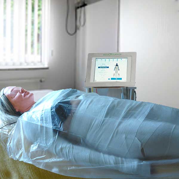
But now there is hope—a new star is rising! Immediate de-acidification is possible if the painful area is rubbed with an alkaline gel and then wrapped in a compression bandage soaked in alkaline saline solution. As you know, saltwater is electrically conductive. This new method achieves its immediate effect through the additional application of frequency-specific microcurrents. The entire bandage acts as an electrode, drawing all positive ions—or protons—to the surface and neutralizing them. Picture it like an electromagnet attracting iron filings.
Admittedly, foam rolling opened up a means of self-treating adhesions in the fascia and hardened muscles. Unfortunately, however, the improvements achieved were often short-lived, requiring one to return to the roller again and again. As long as acidity remains within the tissue, chronic, diffuse pain can hardly be resolved at its root cause. This raises the question: how long do muscles—once de-acidified and stretched using microcurrent therapy—continue to function pain-free? Experience shows that while a single treatment noticeably improves function, the body's acid-base balance is not yet permanently restored. Depending on the duration of the chronic muscle hardening and adhesions, a course of at least three—or up to six—sessions is recommended. Afterward, this improved state should be maintainable through an alkaline-rich diet, ample outdoor physical activity (tailored to one's fitness level), and a positive mindset. And what about those who—just as before—give in to every craving and keep devouring their cake? Well, now we know of a way to remedy those negative consequences in a pleasant manner. Where else is microcurrent therapy helpful? Joint Degeneration Imagine you are on vacation on a volcanic island, eager to finally enjoy some extensive hiking again. On the descent, an increasingly nasty pain begins to develop behind your kneecap. Over the next few days, even climbing stairs becomes painful. Your Achilles tendon feels tender, too. What a shame for such an expensive vacation! Back home, your orthopedist examines your X-ray. Your joint space appears noticeably narrow. The diagnosis is that—because the cartilage behind the kneecap is pressing against the cartilage of the tibia and femur—abrasion is occurring. This can lead to recurring inflammation, which softens and weakens your cartilage. Eventually—once the cartilage has completely worn away, resulting in what is known as "bone-on-bone" contact—the bones will begin to grind directly against one another. This outcome must be prevented. Your orthopedist suggests injecting hyaluronic acid into your knees as a preventive measure. In addition, you receive a prescription for a large supply of ibuprofen. Has a doctor ever explained to you what actually causes the joint space to narrow? What do you think? Is a deficiency in hyaluronic acid—or in anti-inflammatory painkillers—to blame? Most likely not! To be able to bend and extend a joint, you need muscles that span across it. The biceps, for instance, originates near the shoulder and inserts into the forearm. When you contract your biceps, it shortens, and the forearm is lifted. When it relaxes again, its opposing muscle—the antagonist—can easily extend the arm once more. In the thigh, there are several muscles that extend across your knee joint to reach the shinbone. The muscle belly of the *Rectus* muscle, for example, runs across the hip joint, continues down the center of the front of the thigh, and transitions into its tendon just above the kneecap. On its way down to the shinbone, this tendon holds the kneecap in place. A shortened *Rectus* muscle pulls the kneecap tightly against the joint. Also, consider your calf muscles, which extend through the hollow of the knee up toward the thigh. The flexors and extensors normally work together in harmony. However, if unusual physical exertion leads to a buildup of lactic acid (causing muscle soreness), the resulting acidic muscles and adhesions in the fascia will resist stretching—doing so only with pain. The shortened calf muscles pull the lower leg toward the thigh. The rigid muscles of the thigh also contribute to narrowing the joint space. Once the joint has been pulled tight in this manner, the menisci come under intense pressure. Consequently, affected individuals are extremely reluctant to squat down, and their overall mobility becomes severely restricted. Admittedly, this is a purely mechanical approach. However, the realities of clinical practice require such a model in order to arrive at a satisfactory solution. Through microcurrent treatment utilizing alkaline wraps as large-surface electrodes, a significant amount of lactate can be neutralized within just 30 minutes. Prior to this, fascial manipulation and muscle stretching would have been both time-consuming and painful. By the end of this cell-regulating microcurrent treatment, the tissue has been relieved of its high acidic load. The now-improved cellular nourishment—combined with the microcurrent—facilitates the replenishment of ATP. As you may recall, without ATP, muscles are unable to relax. During the final minutes of the session, specific frequencies assist the therapist in performing stretches in a comfortable and pleasant manner. Immediately following the very first session, many patients are able to squat significantly better.
Osteoporosis
In addition to the soft tissues, the skeletal structure also suffers. The reason for this lies in the fact that our body is compelled to maintain the blood pH level stably at around 7.4. Any deviation from this is fatal! When the alkaline minerals supplied through food are no longer sufficient, the body taps into its "treasure chests." These consist not only of the reserves stored in the hair roots (a cause of premature hair loss) but also those within the bones. Almost everyone is familiar with the effects of osteoporosis: a predisposition to spontaneous fractures of the vertebrae or the femoral neck. Orthopedists prohibit heavy lifting or sports that carry a risk of falling—such as cycling. Furthermore, those affected suffer from severe pain. Despite the prescription of expensive medications—such as Vitamin D3—and the intake of mineral supplements, the depletion of bone tissue unfortunately only slows down; the loss of bone density almost invariably continues to progress. In recent years—for instance, at the Mingmen practice in Bad Windsheim—experimental treatments utilizing microcurrent therapy and alkaline body wraps have been employed to combat osteoporosis. The objective was to mitigate the underlying cause—acidosis—through a series of twelve microcurrent sessions, and to stimulate the activity of living bone cells (osteoblasts) using specific frequencies. Applied via a full-body wrap soaked in alkaline saline solution, the bio-electric current was designed to resonate—in a pulsating rhythm—with the osteoblasts. It was hoped that this process would facilitate the regeneration of bone mass by stimulating increased cell division. And lo and behold: the patient became pain-free and resumed jogging. Her quality of life improved enormously. Since bone turnover is a very slow process, bone density was not re-evaluated until six months after the final treatment session. The orthopedist subsequently confirmed that the bone tissue had recovered and that no clinically significant osteoporosis requiring further treatment could be detected. Impaired Wound Healing In numerous studies, microcurrent therapy has proven to be a reliable form of treatment for chronic wound-healing disorders. Advanced tissue acidosis—often accompanied by gelosis (tissue hardening)—is known to compromise the supply of nutrients and oxygen to the affected region. Acting with its innate intelligence, the body attempts to dilute these acids by retaining increased amounts of water. These resulting edemas further lengthen the diffusion distance from the capillaries to the living cells. Consequently, an increasing number of capillaries become occluded by rigid erythrocytes. As a result, normal wound healing becomes severely impeded.
Traditional wound care typically involves cleansing the wound bed and applying firm compression bandages to the legs. A Canadian study published in 2010 in the *Archives of Physical Medicine and Rehabilitation* (Vol. 91, pp. 669–678)—titled "Electrical Stimulation Therapy Increases Rate of Healing of Pressure Ulcers in Community-Dwelling People With Spinal Cord Injury"—monitored the healing progress of two groups of patients suffering from chronic decubitus ulcers (open pressure sores). Both groups received identical standard wound care; however, one group additionally received microcurrent therapy, scheduled for eight hours daily. Unfortunately, patient compliance was poor. Only four patients adhered to the prescribed treatment duration, while the remaining twelve patients underwent treatment for only three hours (± 1.5 hours) per day. Nevertheless, within the three-month observation period, the surface area of the wounds in the microcurrent group shrank by over 70%. In the control group (without microcurrent therapy), the healing success rate stood at 36%. While this represents a respectable result in its own right, it is worth noting that the underlying issue of poor cellular supply to the wounded tissues—caused by acidosis—remained unaddressed in the Canadian study.
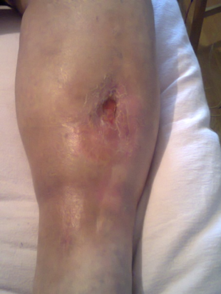
The potential benefits of incorporating alkaline wraps alongside microcurrent therapy are compellingly demonstrated in a case study documented by Birgit Lackert, a practitioner based in St. Wolfgang (84427), Upper Bavaria.
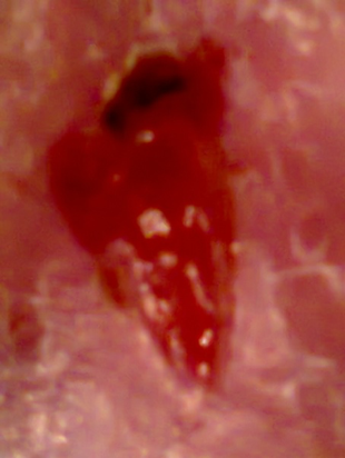
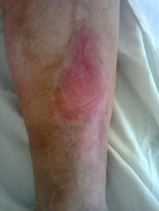
In May 2012, a patient—then 48 years of age—presented at her practice with a prescription for lymphatic drainage. His left tibia presented with a chronic wound resulting from an open comminuted fracture sustained in a traffic accident in 1998. Following surgical intervention, wound healing initially progressed well. In 2004, an ulcer developed that exposed the periosteum of the tibia. Over the course of several years, the patient was prescribed manual lymphatic drainage; however, none of the wound-healing therapies proved effective.
The patient was fit for work but suffered from exercise-induced asthma attacks and became severely overweight (140 kg at a height of 1.78 m). At two-year intervals, the patient was admitted to the trauma clinic for rehabilitation.
From May 6, 2012, onward, the following treatment plan was followed: Wound-healing program utilizing microcurrent therapy, three times per week.